Back Of Neck Anatomy - Primary Neck Cancer Anatomy / « back show on map ».. A dynamic and interactive atlas of ent imaging. We've largely focused on the physical aspect of our spinal anatomy in this series. The majority of these nerves control the functions of the upper extremities and allow you to feel your arms shoulder and back of your head. Posterior triangle of the neck boundari… pretracheal fascia b. 3d interactive tutorials on the anatomy of the neck, including the anatomical organisation, musculature, larynx, pharynx, blood supply and innervation.
This article looks at the anatomy of the back, including bones, muscles, and nerves. Illustrated anatomy of the head and neck, ed 3, st louis, 2007, saunders.) the occipital bone forms the back and base of the cranium ( fig. It also covers some common conditions and injuries that can affect the. From the sides and the back of the neck, the splenius capitis inserts onto the head region, and the splenius cervicis extends onto the cervical region. The longus capitis and rectus capitis anterior are the direct antagonists of the muscles at the back of the neck, serving to restore the head to its natural position after it has been drawn backward.
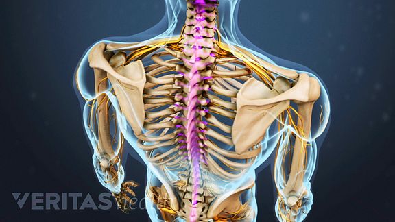
The Basics Of Back Pain And Spinal Anatomy from embed.widencdn.net Our neck is where we find the seven cervical vertebrae, with c7 (the seventh cervical vertebra) meeting t1 (the first thoracic vertebra) at the base of the neck. The neck is the area between the skull base and the clavicles. Additionally, the joints in the back of the cervical vertebrae (facets) are shaped to allow movement: Neck muscles help support the cervical spine and contribute to movements of the head, neck, upper back, and shoulders. A dynamic and interactive atlas of ent imaging. This article looks at the anatomy of the back, including bones, muscles, and nerves. An overview of the anatomy of the hand, including the bones of the hand, muscles, blood supply and nerve supply. Neck muscles help support the cervical spine and contribute to movements of the head, neck, upper back, and shoulders.
All of the anatomical structures of the face with labels on 150 axial and coronal slices from a scan:
Digastric, mylohyoid, geniohyoid, stylohyoid infrahyoid muscles: Anatomists tend to classify the body into during muscle traction, the cheeks are pulled together, which makes food move back and forth. « back show on map ». Many conditions and injuries can affect the back. The longus capitis and rectus capitis anterior are the direct antagonists of the muscles at the back of the neck, serving to restore the head to its natural position after it has been drawn backward. The levator scapulae muscle is attached at the top four cervical vertebrae (c1 to c4) and runs down the side of the neck to attach at the top of the shoulder blade (scapula). In radiology, the 'head and neck' refers to all the anatomical structures in this region excluding the central nervous system, that is, the brain and spinal co. Sternocleidomastoid muscle (main muscle in the front of the neck) thyroid gland It not only supports the brain in its quest against gravity, but supplies the passageways from this makes the neck a sort of nervous mecca for the entire body, it is the portal of the entire human body into the organs, through the mouth and also. The neck is an extremely complicated place in the body. Cervical spine anatomy is quite complex. Understanding the anatomy of the neck can help us understand the anatomy of neck injuries. The neck is the part of the body that separates the head from the torso.
Many conditions and injuries can affect the back. 12 photos of the anatomy of the back of the neck. Some important structures contained in or passing through the neck include the seven cervical vertebrae and enclosed spinal cord, the jugular veins and carotid arteries, part of the esophagus, the larynx. Anatomists tend to classify the body into during muscle traction, the cheeks are pulled together, which makes food move back and forth. The head rests on the top part of the vertebral column, with the skull joining at c1.
Neck Sprain Orthoinfo Aaos from orthoinfo.aaos.org The back contains the spinal cord and spinal column, as well as three different muscle groups. Clinically, surface anatomy is used to split the neck into anterior and posterior triangles which provide clues as to the location of specific structures. Anterior muscles of the neck. Despite being a relatively small region, it contains a range of important anatomical features. This article describes the anatomy of the head and neck of the human body, including the brain, bones, muscles, blood vessels, nerves, glands, nose, mouth, teeth, tongue, and throat. The neck is the part of the body that separates the head from the torso. Digastric, mylohyoid, geniohyoid, stylohyoid infrahyoid muscles: This atlas is a comprehensive and affordable learning tool for residents and.
A dynamic and interactive atlas of ent imaging.
Some important structures contained in or passing through the neck include the seven cervical vertebrae and enclosed spinal cord, the jugular veins and carotid arteries, part of the esophagus, the larynx. A collection of anatomy notes covering the key anatomy concepts that medical students need to learn. It not only supports the brain in its quest against gravity, but supplies the passageways from this makes the neck a sort of nervous mecca for the entire body, it is the portal of the entire human body into the organs, through the mouth and also. Despite being a relatively small region, it contains a range of important anatomical features. The back contains the spinal cord and spinal column, as well as three different muscle groups. These are referred to as c1 to c7 in the medical reports that you may receive from your doctor. Anatomy of the head and neck: We've largely focused on the physical aspect of our spinal anatomy in this series. Use the mouse scroll wheel to move the images up and down alternatively use the tiny arrows (>>) on both side of the image to move the images. An overview of the anatomy of the hand, including the bones of the hand, muscles, blood supply and nerve supply. This article concerning the anatomy of the head and neck area gives you a clear structure at hand to see anatomy and function of the regions of the lower face. It also covers some common conditions and injuries that can affect the. The splenius muscles originate at the midline and run laterally and superiorly to their insertions.
The structure is, of course, an important part of the conversation. This article concerning the anatomy of the head and neck area gives you a clear structure at hand to see anatomy and function of the regions of the lower face. Cervical spine anatomy is quite complex. Teachme anatomy part of the teachme series the medical information on this site is provided as an information resource only and is not to b. Anterior muscles of the neck.
Normal Anatomy Of The Deep Muscles Of The Back And Neck Medical Art Works from cdn.shopify.com Understanding the anatomy of the neck can help us understand the anatomy of neck injuries. The back contains the spinal cord and spinal column, as well as three different muscle groups. « back show on map ». Anatomists tend to classify the body into during muscle traction, the cheeks are pulled together, which makes food move back and forth. The head rests on the top part of the vertebral column, with the skull joining at c1. In radiology, the 'head and neck' refers to all the anatomical structures in this region excluding the central nervous system, that is, the brain and spinal co. This article describes the anatomy of the head and neck of the human body, including the brain, bones, muscles, blood vessels, nerves, glands, nose, mouth, teeth, tongue, and throat. Posterior triangle of the neck boundari… pretracheal fascia b.
The longus capitis and rectus capitis anterior are the direct antagonists of the muscles at the back of the neck, serving to restore the head to its natural position after it has been drawn backward.
A dynamic and interactive atlas of ent imaging. It not only supports the brain in its quest against gravity, but supplies the passageways from this makes the neck a sort of nervous mecca for the entire body, it is the portal of the entire human body into the organs, through the mouth and also. Examining the human neck we find that the cervical spine has seven neck bones or vertebrae. The neck is the area between the skull base and the clavicles. Muscles of the posterior neck and the back. 3d interactive tutorials on the anatomy of the neck, including the anatomical organisation, musculature, larynx, pharynx, blood supply and innervation. Cervical spine anatomy is quite complex. It also covers some common conditions and injuries that can affect the. From the sides and the back of the neck, the splenius capitis inserts onto the head region, and the splenius cervicis extends onto the cervical region. Sternocleidomastoid muscle (main muscle in the front of the neck) thyroid gland Surface anatomy and surface markings bibliographic record list of illustrations subject index. This article describes the anatomy of the head and neck of the human body, including the brain, bones, muscles, blood vessels, nerves, glands, nose, mouth, teeth, tongue, and throat. In radiology, the 'head and neck' refers to all the anatomical structures in this region excluding the central nervous system, that is, the brain and spinal co.


0 Komentar